Elevate Your Practice with Ultrasound Training
- Echo Vet Solutions

- Feb 7
- 4 min read
Updated: Mar 27
Why Ultrasound Matters in Everyday General Practice
Ultrasound has become a cornerstone of modern veterinary diagnostics. It offers a unique window into the body, revealing soft tissue structures that X-rays simply can’t show. This means you can detect issues earlier and with greater accuracy.
For example, imagine you have a dog presenting with vague abdominal pain. An ultrasound scan can help you identify problems like bladder stones, liver abnormalities, or even tumours without invasive procedures. This not only speeds up diagnosis but also reduces stress for your patients.
Moreover, ultrasound is incredibly versatile. You can use it for:
Pregnancy diagnosis and monitoring
Guided biopsies and fluid sampling
Cardiac function evaluation
Musculoskeletal assessments
By incorporating ultrasound into your routine, you’re offering a higher standard of care that benefits both you and your patients.

Essential Skills Every GP Vet Should Develop
If you’re new to ultrasound, the learning curve might seem steep, but don’t worry! With the right approach, you’ll be scanning confidently in no time. Here’s how to get started:
Learn the principles of ultrasound physics, probe types, and image interpretation. This foundation is crucial for accurate diagnosis.
Portable machines are fantastic for on-site use, while more advanced units offer higher resolution for detailed imaging.
Start with simple cases like pregnancy checks or bladder scans. Practice probe placement, image optimisation, and patient handling.
Practical experience is invaluable. Look for courses or workshops that offer live scanning sessions with expert guidance.
Review, discuss cases and use current ultrasound learning resources and guide
Collaborate with colleagues to review images and diagnoses. This helps build confidence and sharpens your skills.
Remember, ultrasound is as much an art as it is a science. Patience and practice are your best allies.
Common Ultrasound Applications in Dogs and Cats
Ultrasound isn’t just for one type of examination. Its applications are broad and continually expanding. Here are some key areas where you can apply your skills:
Abdominal Ultrasound
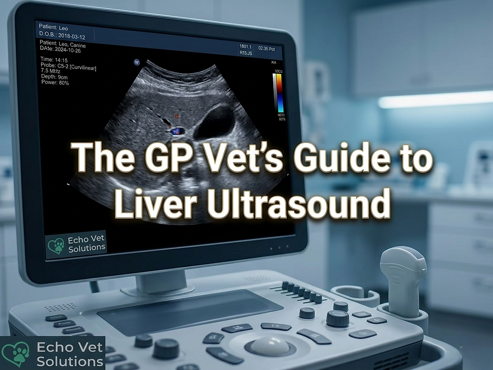
This is the most common use. You can assess organs like the liver, kidneys, spleen, and intestines. It’s especially useful for detecting masses, cysts, or fluid accumulation. You can also examine trickier organs such as adrenals and lymph nodes.
Cardiac Ultrasound (Echocardiography)
Evaluating heart function is critical in many cases. Echocardiography allows you to assess heart chambers, valves, and blood flow. This helps diagnose and manage conditions like cardiomyopathy or valve disease.
Reproductive Ultrasound
From confirming pregnancy to monitoring foetal development, ultrasound is essential in reproductive health. It also aids in diagnosing uterine infections or ovarian cysts.
Musculoskeletal Ultrasound
Though less common, ultrasound can visualise tendons, ligaments, and muscles. This is helpful for diagnosing injuries or inflammation.
Guided Procedures
Ultrasound guidance improves the accuracy of FNA biopsies, fluid taps, and injections, making procedures safer and more effective.
By mastering these applications, you’ll be able to offer comprehensive care tailored to your patients’ needs.

Improving Image Acquisition and Interpretation
Theory is important, but nothing beats hands-on experience. Practical training sessions allow you to:
Handle ultrasound equipment confidently
Recognise normal and abnormal anatomy
Interpret images accurately in a systematic way
Perform guided procedures safely
At Echo Vet Solutions, we focus on delivering practical, on-site training tailored to your clinic’s needs. This means you learn in your own environment, with your own patients, making the experience relevant and immediately applicable.
Training also boosts your confidence. You’ll feel more comfortable discussing findings with clients and making informed decisions. Plus, it’s a great way to stay updated with the latest techniques and technology.
Tips for Integrating Ultrasound into Your Daily Practice
Introducing ultrasound into your routine might seem challenging, but with a few strategies, it becomes second nature:
Start small: Begin with straightforward scans and gradually tackle more complex cases.
Schedule regular practice: Consistency is key to maintaining and improving skills.
Create a reference library: Keep images and notes from previous cases to compare and learn.
Collaborate with colleagues: Share insights and discuss tricky cases to broaden your understanding.
Invest in quality equipment: Reliable machines with good image quality make a huge difference. However, modern machines are able to provide great reliable images without breaking the bank. You do not need top-end equipment to carry out reliable abdominal and cardiac ultrasound exams.
Remember, every scan you perform is a step towards better patient care and professional growth.
How to Build Confidence With Abdominal and Cardiac Scan
If you’re ready to take your ultrasound skills to the next level, consider partnering with experienced trainers who understand your challenges. Echo Vet Solutions is dedicated to helping veterinary professionals across the UK gain confidence and improve their skills through practical, on-site training.
By learning directly in your clinic, you save time and get personalised guidance tailored to your patients and equipment. This hands-on approach ensures you’re not just learning theory but applying it effectively. If you are not sure and you need to find your current ultrasound level, take this quick ultrasound confidence quiz.
Explore how veterinary ultrasound services can transform your practice and enhance patient outcomes. With the right support, you’ll be amazed at what you can achieve!
Tailored In‑Practice Ultrasound Training for Your Team
Now that you’ve got a solid understanding of veterinary ultrasound care, it’s time to put it into action. Whether you’re just starting out or looking to refine your skills, remember that every scan is an opportunity to learn and improve.
Keep practising, seek out training, and don’t hesitate to ask for help when needed. Your patients will thank you for the enhanced care, and you’ll enjoy the satisfaction of mastering a vital diagnostic tool.
Ultrasound is more than just a machine - it’s a bridge to better health and happier patients. Embrace it, and watch your practice thrive!