
From Physics to Practice – Applying Ultrasound Skills in Veterinary Clinics
- Echo Vet Solutions

- Jan 1
- 3 min read
Updated: Mar 27
Introduction
Understanding ultrasound physics and recognising artefacts is crucial. However, the real challenge lies in applying that knowledge consistently in a clinical setting. Every scan requires us to integrate physics, pattern recognition, probe handling, and real-time decision-making—often under time pressure and with patients who don’t always cooperate.
By exploring common clinical scenarios, we highlight how artefacts become diagnostic tools. We also discuss how probe optimisation improves efficiency. Moreover, structured veterinary ultrasound CPD training helps clinicians build confidence in everyday scanning.
Case Examples in Practice
Urinary Calculi

Acoustic shadowing is one of the most reliable sonographic indicators of mineralised material.
What to Look For
A bright, hyperechoic focus with a clean, well-defined distal shadow.
Stones settling dependently within the bladder.
Confirmation in two orthogonal planes.
Why It Matters
Shadowing helps differentiate:
True calculi.
Debris or clots (bright but no clean shadow).
Gas artefacts (reverberation rather than shadowing).
Correct identification prevents misdiagnosis and guides appropriate management of urolithiasis.
Common Pitfalls
Confusing gas reverberation with stones.
Missing small ureteral calculi due to narrow windows.
Assuming all shadows indicate stones (foreign bodies, bone fragments, dystrophic mineralisation).
Practical Tip: Lower the frequency slightly to improve penetration in larger patients or deeper bladder regions.
Fluid Collections and Fine Needle Aspiration (FNA) Biopsy

Acoustic enhancement beneath fluid-filled structures is a key discriminator between cystic and solid lesions.
What to Look For
Anechoic or hypoechoic fluid pocket.
Increased brightness of tissues deep to the fluid.
Smooth, well-defined walls.
Why It Matters
Enhancement helps distinguish:
Simple cysts (strong enhancement, thin walls).
Abscesses (internal echoes but still enhance).
Solid masses (no enhancement; may attenuate instead).
This distinction is crucial in abdominal imaging, where misinterpretation can lead to unnecessary interventions.
Common Pitfalls
Misidentifying a cystic tumour as a simple cyst.
Over-relying on enhancement without assessing wall thickness or vascularity.
Missing small fluid pockets due to suboptimal probe angle.
Practical Tip: Sweep slowly in two planes; enhancement is most obvious when the beam is perpendicular to the fluid interface.
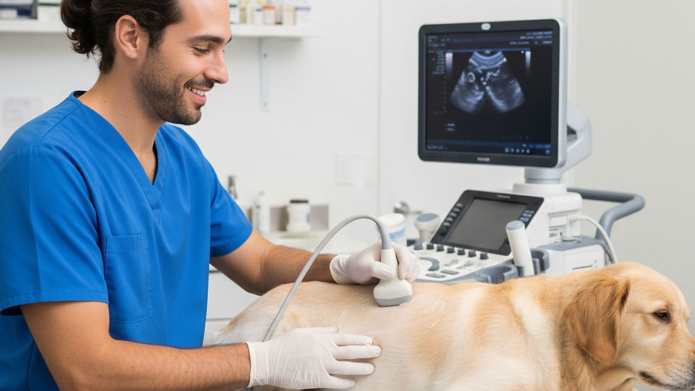
Cardiac Imaging

Cardiac ultrasound is highly sensitive to probe choice, frequency selection, and operator technique.
What to Look For
High frame rate (>80 fps for small animals).
Clear endocardial borders.
Stable long-axis and short-axis views.
Doppler alignment parallel to blood flow.
Why It Matters
Small errors in probe positioning can significantly affect:
Chamber measurements.
Doppler velocities.
Assessment of cardiac function.
Consistency is essential for monitoring disease progression and making evidence-based decisions.
Common Pitfalls
Using too low a frequency → poor resolution.
Tilting instead of sliding → distorted chamber geometry.
Misaligned Doppler → underestimated velocities.
Excessive gain → artefact creation.
Practical Tip: Start with a higher frequency and reduce only if penetration and low frame rate become limiting. Prioritise probe stability—tiny movements have big consequences.
Why CPD Training Matters
Artefact Recognition Prevents Misdiagnosis: Misinterpreting reverberation or mirror artefacts can lead to false positives. It is essential to learn how to distinguish artefacts from pathology.
Probe Optimisation Improves Efficiency: Selecting the right frequency and transducer type saves time in busy clinics and enhances diagnostic accuracy.
Hands-On Practice Builds Confidence: Small-group, practical sessions consistently allow us to refine skills in a supportive environment.
The Importance of Continuous Learning
Continuous professional development (CPD) is vital in our field. As veterinary professionals, we must stay updated with the latest techniques and technologies. This ensures we provide the best care for our patients. Engaging in CPD training not only enhances our skills but also boosts our confidence in applying ultrasound in clinical practice.
Benefits of Practical Training
Practical training allows us to apply theoretical knowledge in real-world scenarios. This hands-on experience is invaluable. It helps us understand the nuances of ultrasound imaging better. Moreover, it fosters a collaborative learning environment where we can share experiences and insights with peers.
Building a Community of Practice
Participating in CPD training creates a community of practice. We can connect with fellow veterinary professionals who share similar challenges and successes. This network provides support, encouragement, and opportunities for collaboration. Together, we can improve our skills and enhance patient care.
Conclusion
Ultrasound becomes a powerful diagnostic tool only when we combine a solid understanding of physics with confident, practical scanning skills. Recognising artefacts, optimising probe settings, and interpreting images consistently are competencies that develop through structured practice.
We support veterinary teams in building these skills through tailored in-practice CPD training, using your clinic’s own equipment to ensure every session is directly relevant to your workflow. Our onsite courses cover echocardiography, abdominal ultrasound, and emergency scanning, giving your team practical skills they can apply immediately.
For deeper study, you can explore curated reference papers via our Resources for Vets page. By combining physics, artefact recognition, and hands-on scanning, we help your team turn theory into confidence, and confidence into better patient outcomes.

