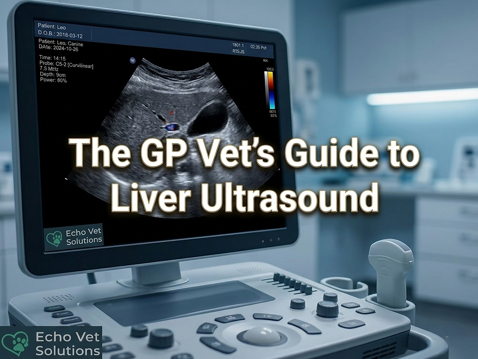
Small Animal Abdominal Ultrasonography: Getting the Best Out of Your Ultrasound Images
- Echo Vet Solutions

- Dec 10, 2025
- 2 min read
Updated: Jan 4
Abdominal ultrasound is one of the most versatile diagnostic tools in veterinary medicine. For small animals, abdominal ultrasonography provides a safe, non-invasive way to evaluate internal organs and guide clinical decisions. But producing consistently clear, diagnostic-quality images requires more than just switching on the machine. It’s about understanding the controls, preparing the patient properly, and applying a systematic scanning technique.

Why Image Optimization Matters
Even the most advanced ultrasound machine cannot compensate for poor technique. Image optimization ensures that subtle lesions are visible, artefacts are minimized, and diagnostic accuracy is maximized. For veterinary teams, this knowledge builds confidence and prevents misinterpretation.
Key Machine Controls
To achieve reliable abdominal ultrasound scans, practitioners should be familiar with these essential settings:
Probe selection – Microconvex transducers are often ideal for abdominal imaging.
Frequency – Higher frequencies give sharper resolution but less depth; lower frequencies penetrate deeper.
Depth and focal zone – Adjust depth to the organ of interest and place the focal zone just beneath it.
Gain and TGC – Balance brightness across near and far fields to avoid washed-out or overly dark images.
Dynamic range – Mid-range contrast helps distinguish subtle tissue differences.
Presets – Save optimized settings for consistency across examinations.

Patient Preparation
Clipping fur, applying alcohol, and using plenty of gel improve probe contact and reduce artefacts. A calm, medium-sized dog makes an excellent practice patient when learning these techniques.
Scanning Technique
Hold the transducer perpendicular to the skin for accurate beam orientation.
Examine abnormalities in two planes (long and short axis).
Follow a systematic approach: liver, spleen, kidneys, bladder, intestines.

Practical Tips
Experiment with each control to understand its impact.
Start with mid-range settings and adjust gradually.
Document patient data carefully for image storage and retrieval.
Practice on cooperative patients before tackling complex cases.
Conclusion
Small animal abdominal ultrasonography is a skill that blends technical knowledge with hands-on practice. By mastering machine controls, patient preparation, and scanning technique, veterinary teams can consistently produce diagnostic-quality images. Attention to detail transforms ultrasound into a powerful window into animal health.
Want to deepen your understanding of small animal abdominal ultrasonography? Discover our essential and advanced veterinary ultrasound training courses and gain practical skills in image optimization, patient preparation, and systematic scanning techniques. With the right support, every veterinary team can build confidence step by step, and you can explore our resources for vets to continue learning and applying these skills in practice.